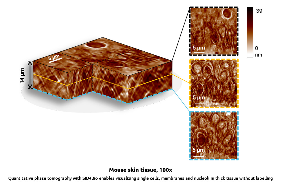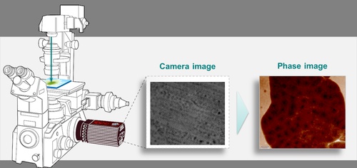
The Phasics innovative technique enables fast protocols for tissue imaging with no staining nor labelling. The resulting images are artefact-free and offer high contrast to observe tissue structures including cells and fibers.
It can be applied to:
- Patient clinical follow-up: biopsy, anatomic pathology including digital pathology and rapid intraoperative examinations
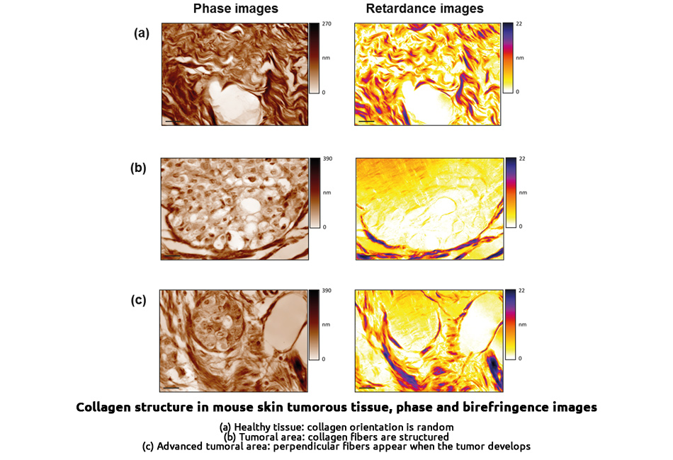
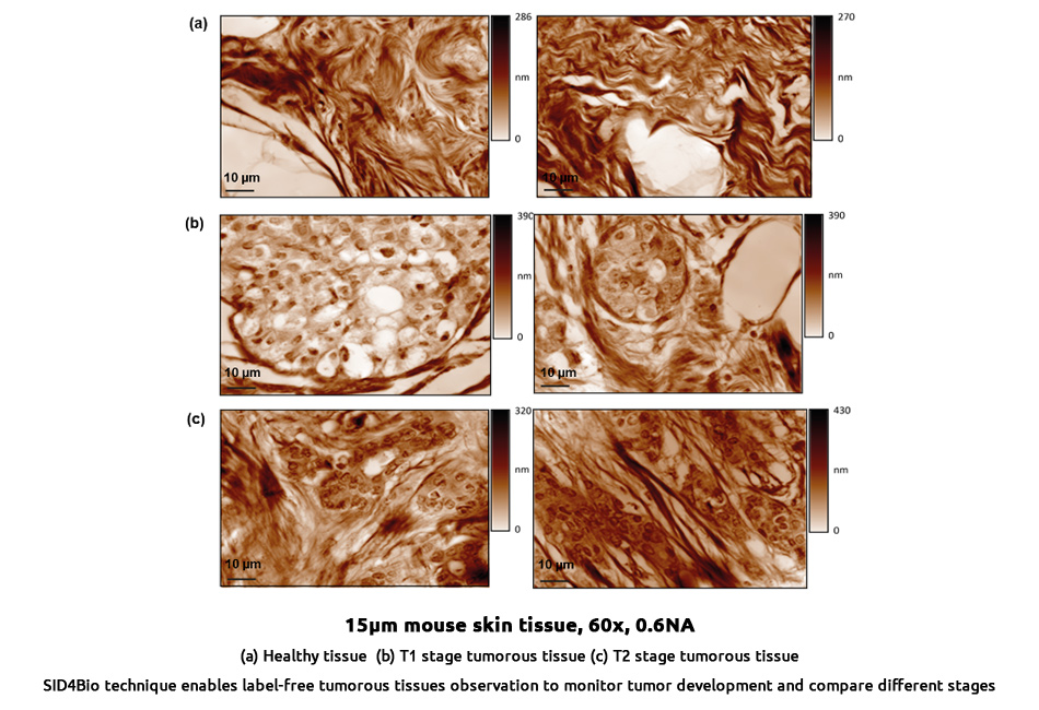
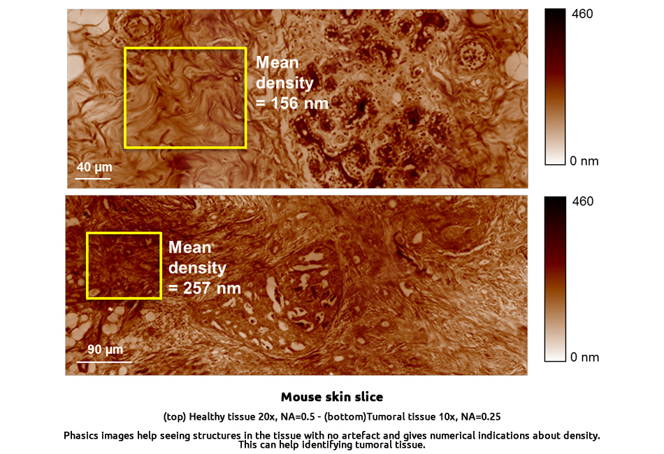
- Cancer diagnosis and monitoring using collagen orientation in tumors
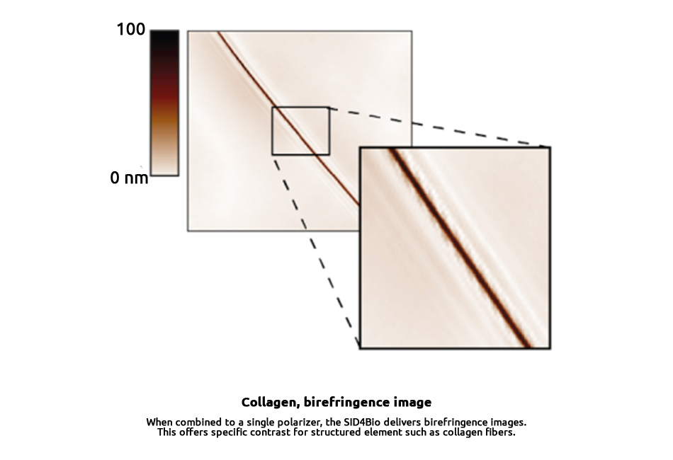
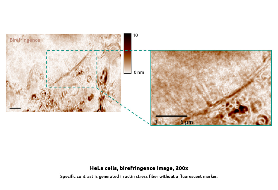
- Fiber detection and identification
- Tissue morphology study